Thank you for visiting nature.com. You are using a browser version with limited support for CSS. To obtain
the best experience, we recommend you use a more up to date browser (or turn off compatibility mode in
Internet Explorer). In the meantime, to ensure continued support, we are displaying the site without styles
and JavaScript.
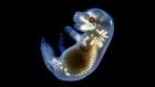
Human neurons made from stem cells (green) two weeks after being transplanted into a mouse brain. Credit: Pierre Vanderhaeghen
In a darkened room in a laboratory in London, a group of students and researchers watch a clump of human brain cells settle into their new home: a living mouse brain. On a computer monitor next to a microscope, the human cells light up in flashes of simultaneous activity. Over time, the cells sprout new connections a few centimetres long, and form networks with each other. It’s captivating viewing for his students, says Vincenzo De Paola, who runs the lab at Imperial College London. “It’s all they want to do. I can’t tear them away,” he says.
They have front-row seats to an unusual show. De Paola’s group is one of just a handful of labs able to study human neural cells at work in a live, developing brain — a system that is otherwise largely off limits for both ethical and technical reasons. “We cannot study these processes as they unfold in a fetal human brain,” he says. “Instead, we wanted to watch human cortical neurons mature and form active networks in a live animal.”
De Paola’s system is a specialized type of neural chimaera — an area of research that has expanded immensely in the past five years, sparking a debate about the ethics of blending human and animal brain tissue. Proponents say that such systems are necessary to manipulate live human neurons and are already yielding important insights into health and disease. For example, using neural chimaeras, scientists have found differences in how neurons develop and behave in Down’s syndrome and Alzheimer’s disease.
But others warn that such chimaeras represent an ethical grey zone, because of the potential to blur the line between humans and other animals, or to recapitulate human-like perception or cognition in an animal. Some researchers say these kinds of chimaeras should only be used if no other cell or animal model is appropriate. “Is this a really good model for answering a scientific question or are we pushing boundaries for the sake of it?” asks Naomi Moris, a developmental biologist at the Francis Crick Institute in London. Ethicists are asking at what point a collection of human neurons in another animal’s brain embodies something that deserves a unique moral status.
Although research using chimaeras — entities made up of cells from different organisms or species — has been going on for decades, these neural chimaeras broach new ethical territory. A 2021 special report on neural chimaera research by the US National Academies of Science, Engineering and Medicine (see go.nature.com/3pii9q5) flagged issues such as the possibility of endowing animals with new cognitive abilities or human disease symptoms that could be distressing. The committee advised that although current regulation of stem-cell and animal research was adequate, the field should be kept under close surveillance. The committee also encouraged the use of pilot studies and close monitoring of animals to identify any new or unusual behaviours.
There will be plenty for regulators to keep an eye on. Researchers are starting to consider going beyond transplanting a few isolated cells to creating chimeric animals with human brain regions. Studies that transplanted human brain stem cells into monkeys’ brains helped to launch a 2018 clinical trial testing whether human brain stem-cell transplants can treat Parkinson’s disease. In 2019, Japan reversed a ban on government funding for research using human–animal chimeric embryos. In many countries, including the United States and the United Kingdom, research that mixes human brain cells or tissue with another animal’s brain is legally allowed, and can be government-funded with an extra layer of review. The United States prohibits government funding for human–animal chimeric embryo research.
Observers expect the field to be fast-moving. “We know it will be quickly evolving,” says Insoo Hyun, director of research ethics at Harvard Medical School in Boston, Massachusetts.
A research mainstay
The history of biology abounds with chimaeras. Starting in the early 1900s, embryologists cut and pasted together pieces of embryo from different animal species — merging a chicken with a quail, for instance, to work out where developmental signals originated, says Ali Brivanlou, a developmental biologist at Rockefeller University in New York City.
Researchers have also been introducing human elements such as organs, cells or genes into other animals for decades. Often, the rationale is to better understand how biological systems are working, says Brivanlou, or to find treatments for disease. Cancer researchers routinely transplant human tumours into mice, and since the late 1980s scientists have created mice with human immune systems1.
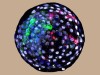
Scientists transplanted a human organoid (bright green) into a mouse brain to study how neurons behave in the context of health and disease.Credit: Abed Mansour
Another motivation is more utilitarian: to find ways to grow human-compatible organs in animals to alleviate the shortage of organs for transplantation. In the past few months, researchers have transplanted genetically modified pig kidneys and a pig heart into humans.
But transplants of human neurons that survive long-term have only been created in the past decade. In 2013, Pierre Vanderhaeghen, a neuroscientist then at the Université Libre de Bruxelles, and his colleagues perfected the delicate process2 of growing human neurons from stem cells to the point that they would flourish — but not grow uncontrolled — in the mouse brain when transplanted.
Scientists use two types of human stem cell to make neurons for chimaeras: either embryonic stem cells (ES cells), which are originally derived from embryos, or induced pluripotent stem cells (iPS cells), which are derived from adult cells that are reprogrammed to an embryonic-like state. Both types have the potential to become any tissue in the body and can be directed to grow into neurons. “Cells derived from human pluripotent stem cells are a lot more plastic than other cells transplanted in the past,” which could result in better integration of the human cells, says Hyun.
In 2016, the US National Institutes of Health proposed lifting its funding moratorium on research using animal embryos containing human cells. Comments from the public, largely expressing opposition, flooded into the agency. (The funding ban remains in place.) But a 2020 survey of 430 people in the United States found that 59% supported human–pig chimeric embryo research designed to produce human tissues in pigs3.
Pigs with human kidney or liver tissue are one thing; neural tissue might not be so acceptable. “It is the brain that people associate with moral status,” says Hyun. Although researchers say none of the research comes close to producing human-like cognition in an animal, this association is making them reconsider — at what point does an animal’s brain become too human-like for society’s comfort?
Mingling brain cells
In the past five years, researchers have developed several ways to make neural chimaeras. They vary in complexity from transplanting single human neurons or a chunk of cultured brain tissue, to combining embryos from two species to try to produce chimeric brain tissue from scratch.
The most minimal way to get a rare glimpse of human neurons at play is to transplant just a few cells at a time. Vanderhaeghen’s group, now at the Flemish Institute of Biotechnology (VIB)–Catholic University of Leuven (KU Leuven) Center for Brain & Disease Research in Belgium, has been doing this with pyramidal neurons — the most abundant type in the human cortex — grown in a dish from ES cells. They wondered how the cells might wire up over longer timescales in a living animal. “We wanted to know how those neurons trained in the dish would act in the battlefield of the brain,” says Vanderhaeghen.
His group, working with Vincent Bonin’s team at VIB Neuro-Electronics Research Flanders in Leuven, transplanted a soup of human neurons that integrated as single cells, rather than as a clump, into the cortex of a newborn mouse4.
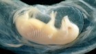
Human brain cells and their nuclei two months after being transplanted into a mouse brain.Credit: Raquel Real, Manuel Peter, Rick Livesey and Vincenzo De Paola
The human neurons took their customary time to mature, between 6 and 12 months compared with 5 weeks for their mouse neuron neighbours. Even in the environment of the mouse brain, they stuck to their lengthy timeline, says Vanderhaeghen. “This suggested that this prolonged developmental timing is encoded intrinsically, in the neurons themselves.”
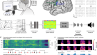
The team found that the human neurons developed normally, integrated into and functioned within the mouse’s visual circuit, responding just as mouse cells did to visual stimuli, such as moving black and white bars. That the human neurons settled into a foreign brain and worked normally was surprising — and it hints that cell transplants might be used to repair damaged brain circuits in the future.
“We expected some connectivity, but we were quite stunned at how specific the responses were,” says Bonin. “There are a million ways this could have failed.”
The team has also transplanted healthy human neurons into the brains of mice with a genetic predisposition to Alzheimer’s disease. The work5 showed that the human neurons degenerate in the diseased brain, whereas the mouse neurons remained alive. This not only confirmed that human neurons are particularly vulnerable to Alzheimer’s disease, but also gave researchers a way to watch what happens to human neurons in a living diseased brain.
De Paola, who also runs a group at Duke–NUS Medical School in Singapore, studies how human neurons connect with one another and how this is disrupted in developmental disorders. His group grafted pyramidal neurons made from human iPS cells into the somatosensory cortex of adult mice6.
In contrast to Vanderhaeghen’s transfers, these transplants grew into dense micrografts of human tissue in the mouse brain and survived until the experiment ended after five months. “We were surprised at how much growth there was, it was a massive network,” says De Paola. “Well, ‘massive’ is relative — it was about the size of a large lentil.”
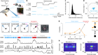
The grafted cells kept mostly to themselves — more than 90% of the connections were human to human — but they did send out projections to other parts of the mouse cortex, and received a few projections, blood vessels and immune cells from the mouse brain, he says. Those supports allowed the chunk of tissue to keep developing for five months, playing out the behaviour normally expected in a developing human fetal brain — pruning neuronal branches and connections, and starting to fire in coordinated waves.
De Paola’s team did the same transplant experiment using neurons made from cells from people with Down’s syndrome6. They found that these neurons formed less dynamic networks, with lower neural activity — but it’s unclear what relationship, if any, exists between the two features. The team is exploring that next. “We can do that experiment in this model. Obviously, we cannot do it in a human adult or fetal brain,” De Paola says.
Could these groups’ transplants somehow change the mouse’s visual or sensory perception to a more human version? Neither team has tested cognition or behaviour in the transplanted mice, but all report that the mice generally behaved like their non-transplanted peers. Both De Paola and Vanderhaeghen are sceptical that the limited numbers of human neurons and connections could change a mouse’s outlook. “I don’t think stimulating even a few thousand human cells would drive human behaviour or perception,” says Vanderhaeghen. But he and De Paola think that those working in the field should try to determine at what point that might change.
Long-lived organoids
A major advance in the study of human brain tissue in the laboratory has been the rise of brain organoids, self-organizing structures formed when brain stem cells are grown in 3D culture.
Brain organoids have become increasingly intricate since they were first created in 2013 by Madeline Lancaster and Jürgen Knoblich7. Some researchers have even stitched multiple organoids together into ‘assembloids’.
Organoids are complex enough to be a good way to ask many questions about the human brain, but even assembloids are still far from the complexity of the real thing, says Sergiu Pasca, a neuroscientist at Stanford University in California. That’s because they lack sensory input, blood vessels, immune and support cells, and don’t receive feedback, he says. Plus, once the structures grow beyond 3–4 millimetres in size, the cells in the middle die owing to lack of nutrients from the cell-culture broth. It can be hard to support their growth beyond a couple of months.
To overcome these limitations, neuroscientists have begun to transplant organoids into an animal’s brain to more closely model the complexity of human brain circuits and how they go awry in disease.
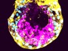
Organoids, such as this one made from human neurons, have been transplanted into animal brains to study how neurons connect and communicate.Credit: Ilaria Chiaradia
Neuroscientist Rusty Gage’s group at the Salk Institute for Biological Studies in La Jolla has succeeded in transplanting human organoids into mouse brains and keeping them alive for up to 11 months, nearly the mouse’s entire lifespan8. Using this system, they have unpublished results showing that the human neurons mature from an embryonic-like condition to a more complex state akin to neurons in an infant, and eventually show characteristics of adult neurons. The human brain tissue integrated into the mouse brain, grew blood vessels, matured and responded to stimuli, and even formed sparse, but working, connections with mouse neurons.
Abed Mansour, who established the organoid transplants as a postdoctoral fellow working with Gage, says the system has advantages for studying what happens to neurons in neurodegenerative disorders such as Alzheimer’s disease. Human neurons in organoid transplants send long projections into the host brain. “This might become an excellent system to ask how this process differs between healthy human neurons and disease-affected neurons,” says Mansour, who now leads his own group at the Institute for Medical Research at the Hebrew University of Jerusalem.
Gage’s group now plans to transplant brain organoids made from the cells of people with Alzheimer’s disease into healthy mouse brains and, conversely, healthy human organoids into mouse brains that mimic Alzheimer’s symptoms. The aim is to tease apart which cell types — the neurons themselves, or other brain cells such as astrocytes — contribute to the inflammation seen in the disease.
“For the first time, we are able to monitor living human brain tissue in a disease context,” Gage says. One day, he says, this research could lead to personalized organoid transplants that replace diseased or injured brain tissue.
For Lancaster, now a developmental biologist at the MRC Laboratory of Molecular Biology in Cambridge, UK, organoid transplantation has its place, but she urges researchers to examine closely the animal experiments they are doing and make sure they are justified. “We need to be careful as researchers — this is such a hot field with a lot of papers being published,” she says.
As for the ethical status of organoids, when in the dish they are essentially considered to be a fancy 3D cell culture. Lancaster, Gage and others do not consider them to be capable of human perception, sensation or cognition. And Gage says that the transplanted organoids do not integrate well enough to confer any meaningful ‘human-ness’ either.
Enjoying our latest content?
Login or create an account to continue
Access the most recent journalism from Nature's award-winning team
Explore the latest features & opinion covering groundbreaking research


 What’s next for lab-grown human embryos?
 Can lab-grown brains become conscious?
 First monkey–human embryos reignite debate over hybrid animals
 The rise of the assembloid